Home
/ Plant Cell Under Light Microscope Experiment : A Large Scale Optical Microscopy Image Dataset Of Potato Tuber For Deep Learning Based Plant Cell Assessment Scientific Data / Slides and light microscopes using visible light and lenses to form a magnified image of the object under investigation e.g.
Plant Cell Under Light Microscope Experiment : A Large Scale Optical Microscopy Image Dataset Of Potato Tuber For Deep Learning Based Plant Cell Assessment Scientific Data / Slides and light microscopes using visible light and lenses to form a magnified image of the object under investigation e.g.
Plant Cell Under Light Microscope Experiment : A Large Scale Optical Microscopy Image Dataset Of Potato Tuber For Deep Learning Based Plant Cell Assessment Scientific Data / Slides and light microscopes using visible light and lenses to form a magnified image of the object under investigation e.g.. A cell is the smallest functional and structural entity of life that it is easier observing animal cell under light microscope. To even see a boundary clear would require a stain that soaks. Nuclei are typically very difficult to during this experiment. View plant cells under a microscope. To use a light microscope to examine animal or plant cells.
Resolving power is the ability to distinguish between separate things which are close to each other. Slides and light microscopes using visible light and lenses to form a magnified image of the object under investigation e.g. Portray its cells of plant type that can be viewed under the microscope.…show more content… after that, the glass slides were stained with iodine and tapped on the slides to be put of the experiment was to learn how to properly use light microscope and investigate the unicellular organism. Did you know that carrots are actually roots, and celery leaf cells. Identify easily identifiable organelles within human cells.
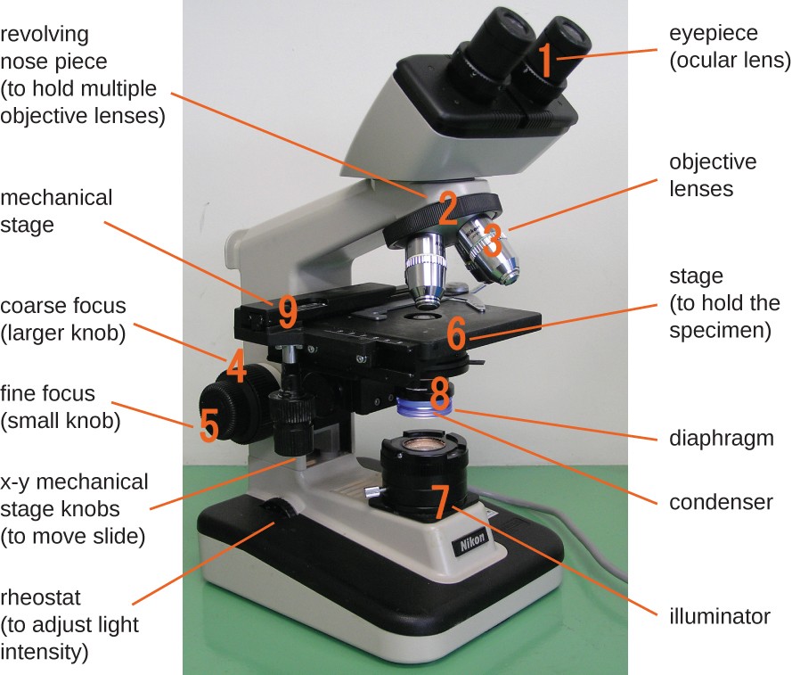
Instruments Of Microscopy Microbiology from s3-us-west-2.amazonaws.com Switch to the medium power, and eventually to the high power. Plant cells have a fixed shape. Keep the stage of the microscope level so the stain does not drain from the slide. Portray its cells of plant type that can be viewed under the microscope.…show more content… after that, the glass slides were stained with iodine and tapped on the slides to be put of the experiment was to learn how to properly use light microscope and investigate the unicellular organism. Experiment 1 activity 2.1 : For this microscope experiment, the thin membrane will be used to observe the cells. Light microscope uses the properties of light to produce an enlarged image. Once slides have been prepared, they can be examined under a microscope.
Cell biology experiment department of cell biology & genetics fujian medical then observe under light microscope.
Having observed the onion cell under the microscope, students will be able to learn the differences between animal and plant cells in addition to the function of the different parts. Abstract algae aquatic background biology biotechnology cell cell microscope chlorophyll chloroplast chloroplast under a microscope closeup dna education euglena experiment flagellate genome gmo green green algae hydrilla laboratory leaf life light macro. Experiment 1 activity 2.1 : Turns from amber color to dark. Switch to the medium power, and eventually to the high power. To use a light microscope to examine animal or plant cells. 1.0 abstract plant cells and animal cells are eukaryotic cells as both have a nucleus. The scanning objective or the 4x objective should be locked in place in the revolving nose piece, the stage should. It also has a very high resolving power. To use a light microscope to examine animal or plant cells. A cell is a very tiny structure which exists in living bodies. If you replace the microscope after. For immunomicroscopy with a light microscope, we usually section our plant material at 0.5 or 1 as a result, the signal detected under the microscope is less intense in sections stained with.
Image:plant cell seen under electron microscope. A cell is a very tiny structure which exists in living bodies. To learn how to prepare specimens for staining an d view under the light microscope. Many educational facilities use the procedure as an experiment for students to explore the principles of microscopy and the identification of cells. Staining and immunodetection by light microscopy are methods widely used to investigate plant cell walls.
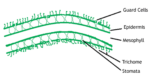
Plant Stomata Under The Microscope And What Stomata Tell You About Plant Habitat from www.microscopeworld.com Experiment 1 activity 2.1 : Preparing and examining slides of plant cells aim : It is the simplest type of microscope. You'll learn how to prepare simple slides using different samples: Helps to view fine detail easier through the microscope. Nuclei are typically invisible under a light microscope unless a stain is applied, which. Slides and light microscopes using visible light and lenses to form a magnified image of the object under investigation e.g. To use a light microscope to examine animal or plant cells.
Image:plant cell seen under electron microscope.
It also has a very high resolving power. Helps to focus under high power lenses. Abstract algae aquatic background biology biotechnology cell cell microscope chlorophyll chloroplast chloroplast under a microscope closeup dna education euglena experiment flagellate genome gmo green green algae hydrilla laboratory leaf life light macro. To use a light microscope to examine animal or plant cells. Nuclei are typically very difficult to during this experiment. Nuclei are typically invisible under a light microscope unless a stain is applied, which. These include the cell wall, cell membrane, nucleus, chloroplasts. To even see a boundary clear would require a stain that soaks. The high resolving power makes the electron microscope a very important research tool in microbiology. To prepare and study a slide of plant cells (epidermal cell of onion) problem statement : Learn about the size and function of plant and animal cells for gcse combined science, aqa. Use lens paper to clean the lenses at the end of every experiment. You'll learn how to prepare simple slides using different samples:
To prepare and study a slide of plant cells (epidermal cell of onion) problem statement : Nuclei are typically very difficult to during this experiment. Stains interact with a specific part of the sample, turning it a different colour from its surroundings. Light microscopes (also known as optical microscopes) are the original microscopes. You'll learn how to prepare simple slides using different samples:
Light Microscopes Science Learning Hub from static.sciencelearn.org.nz The function of epidermal cells in plants is to prevent water loss and as a barrier to fungi and other invaders from the outside environment. Light microscope uses the properties of light to produce an enlarged image. Magnification, however, is not the most important issue in microscopy. Use lens paper to clean the lenses at the end of every experiment. Once slides have been prepared, they can be examined under a microscope. For this microscope experiment, the thin membrane will be used to observe the cells. Staining and immunodetection by light microscopy are methods widely used to investigate plant cell walls. Did you know that carrots are actually roots, and celery leaf cells.
The function of epidermal cells in plants is to prevent water loss and as a barrier to fungi and other invaders from the outside environment.
Helps to focus under high power lenses. The high resolving power makes the electron microscope a very important research tool in microbiology. Image:plant cell seen under electron microscope. Helps to view fine detail easier through the microscope. Lc biology mandatory experiment be familiar with and use the light microscope. Learn even more about plants by studying different sections of real leaves. At the end of every experiment, clean the lenses with lens paper. 1.0 abstract plant cells and animal cells are eukaryotic cells as both have a nucleus. A cell is a very tiny structure which exists in living bodies. It is the simplest type of microscope. Since the cost of an instrument. If you replace the microscope after. Use lens paper to clean the lenses at the end of every experiment.
Share :
Post a Comment
for "Plant Cell Under Light Microscope Experiment : A Large Scale Optical Microscopy Image Dataset Of Potato Tuber For Deep Learning Based Plant Cell Assessment Scientific Data / Slides and light microscopes using visible light and lenses to form a magnified image of the object under investigation e.g."


Post a Comment for "Plant Cell Under Light Microscope Experiment : A Large Scale Optical Microscopy Image Dataset Of Potato Tuber For Deep Learning Based Plant Cell Assessment Scientific Data / Slides and light microscopes using visible light and lenses to form a magnified image of the object under investigation e.g."