Home
/ Plant Cell Structure Under Microscope - Chloroplast Under A Microscope Chloroplasts Stock Footage Video 100 Royalty Free 1028546699 Shutterstock / This gives rise to its name of rough endoplasmic reticulum.
Plant Cell Structure Under Microscope - Chloroplast Under A Microscope Chloroplasts Stock Footage Video 100 Royalty Free 1028546699 Shutterstock / This gives rise to its name of rough endoplasmic reticulum.
Plant Cell Structure Under Microscope - Chloroplast Under A Microscope Chloroplasts Stock Footage Video 100 Royalty Free 1028546699 Shutterstock / This gives rise to its name of rough endoplasmic reticulum.. 8 pictures of plant cells under a microscope. 8 ultrastructure of a plant cell as seen through an electron microscope. 9 pupil activity cell structure read through the information on each of the organelles as you colour them in follow the guidance on colouring them in given at the bottom of the page this works on the theory that whilst you are. A plant cell than can be used in this activity to replace the onion cell is. This gives rise to its name of rough endoplasmic reticulum.
We say cells are microscopic because they can only be seen under a microscope. Your plant cells under microscope stock images are ready. Image:plant cell seen under electron microscope. These are both specific types of cells, and. Examining specimens under a good microscope enables us to study these cellular structures and investigate their biological functions.
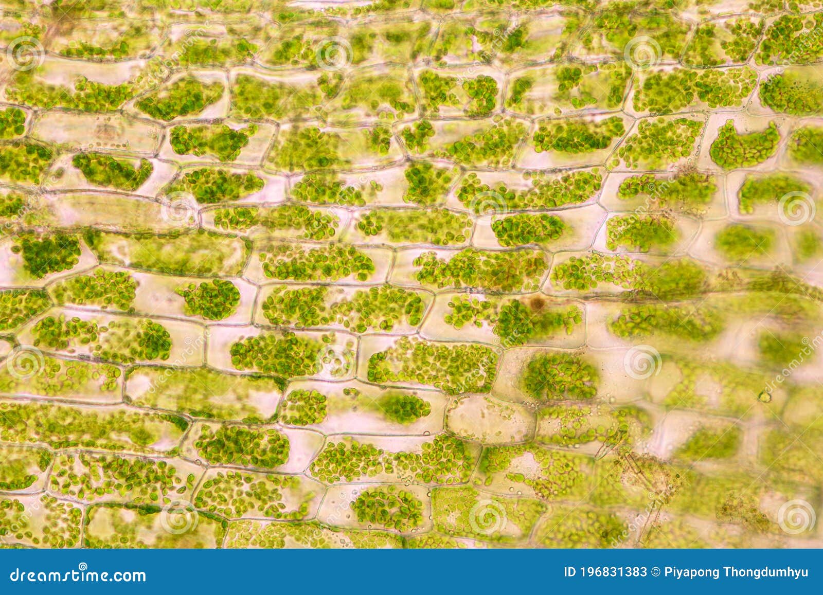
Cell Structure Hydrilla View Of The Leaf Surface Showing Plant Cells Under The Microscope Stock Image Image Of Micro Aquatic 196831383 from thumbs.dreamstime.com Image:plant cell seen under electron microscope. Dreamstime is the world`s largest stock photography community. A micrograph is a photo or digital image taken through a microscope to show a magnified image of a specimen. Epidermal cells include several types of cells that make up the epidermis of plants. The type of cell and the structure of cells. Differences between a plant and an animal cell. The diagram is very clear, and labeled the animal cell is more fluid or elastic or malleable in structure; Learn vocabulary, terms and more with flashcards structure:
Here's a photo of a plant cell under an electron microscope.
This gives rise to its name of rough endoplasmic reticulum. We say cells are microscopic because they can only be seen under a microscope. There are various tasks done by a cell to complete them as the cell is the basic purposeful and. Now that we have looked at the basic structures and functions of the organelles in a cell, you would have noticed that there are key differences between plant and animal cells. The plant cell as more rigid and stiff walls. Their distinctive features include primary cell walls containing cellulose, hemicelluloses and pectin, the presence of plastids with the capability to perform photosynthesis and store starch. Plant cells have cell walls, one large vacuole per cell, and chloroplasts, while animal cells will have a cell membrane only. The first microscopes were composed of a single lens just like a magnifying glass. Guide to plant anatomy and biology using microscope premade slide sets. Robert hooke in 1665 first discovered plant cell. The term 'cell' was coined to describe the small walled units that were observed in the sections of bottle cork under simple they lack any internal structure and contain hydrolytic enzymes. Learn vocabulary, terms and more with flashcards structure: Dreamstime is the world`s largest stock photography community.
The term 'cell' was coined to describe the small walled units that were observed in the sections of bottle cork under simple they lack any internal structure and contain hydrolytic enzymes. When viewed under the microscope, it is possible to view the cell nucleus, a very thin layer of cytoplasm that can be seen in. Plant cells have a regular shape and structure and keep their shape easily. We say cells are microscopic because they can only be seen under a microscope. Examining plant cells under the microscope.
Cell Structure Hydrilla View Of The Leaf Surface Showing Plant Cells Under The Microscope For Classroom Education 411280030 Larastock from st4.depositphotos.com Animal cells also have a because only plant cells perform photosynthesis, chloroplasts are found only in plant cells. Their distinctive features include primary cell walls containing cellulose, hemicelluloses and pectin, the presence of plastids with the capability to perform photosynthesis and store starch. Examining specimens under a good microscope enables us to study these cellular structures and investigate their biological functions. A plant cell than can be used in this activity to replace the onion cell is. Plant cells have cell walls, one large vacuole per cell, and chloroplasts, while animal cells will have a cell membrane only. This gives rise to its name of rough endoplasmic reticulum. A microscope is an important instrument for studying cells e.g. Plant cells are eukaryotic cells present in green plants, photosynthetic eukaryotes of the kingdom plantae.
We say cells are microscopic because they can only be seen under a microscope.
Glass was developed by the romans in the first century. Differences between a plant and an animal cell. Resolving power is the ability to distinguish between separate things which through the electron microscope, very fine details of the cell can be observed. Your plant cells under microscope stock images are ready. Here's a diagram of a plant cell: When viewed under the microscope, it is possible to view the cell nucleus, a very thin layer of cytoplasm that can be seen in. Animal cells also have a because only plant cells perform photosynthesis, chloroplasts are found only in plant cells. Start studying cell structure & microscopes. Plant cells have cell walls, one large vacuole per cell, and chloroplasts, while animal cells will have a cell membrane only. Structure and functions of the cell organelles. This gives rise to its name of rough endoplasmic reticulum. Light and electron microscopes allow us to see inside cells. The type of cell and the structure of cells.
Plant cells are eukaryotic cells present in green plants, photosynthetic eukaryotes of the kingdom plantae. A plant cell than can be used in this activity to replace the onion cell is. Guide to plant anatomy and biology using microscope premade slide sets. Robert hooke was the first cytologist to identify cells under his microscope in 1665. The diagram is very clear, and labeled the animal cell is more fluid or elastic or malleable in structure;
Botany Online Cells And Tissues What Is Viewed In A Microscope from www1.biologie.uni-hamburg.de These are both specific types of cells, and. Plant, animal and bacterial cells have smaller components each how have light microscopes developed? Plant cells have a regular shape and structure and keep their shape easily. The diagram is very clear, and labeled the animal cell is more fluid or elastic or malleable in structure; To examine a specimen like plant or animal cells under a microscope you need to prepare microscope slide. This gives rise to its name of rough endoplasmic reticulum. The colour of the nucleus that is stained with iodine solution is. Membrane bound vesicle containing digestive enzymes.
A micrograph is a photo or digital image taken through a microscope to show a magnified image of a specimen.
A microscope is an important instrument for studying cells e.g. Examining specimens under a good microscope enables us to study these cellular structures and investigate their biological functions. The colour of the nucleus that is stained with iodine solution is. This means a cell does various tasks that it was designed to accomplish. They are green in color under a microscope because they. Definition, function, structure and microscopy. Plant cells have a regular shape and structure and keep their shape easily. Plant cells have a fixed shape. 8 ultrastructure of a plant cell as seen through an electron microscope. The first microscopes were composed of a single lens just like a magnifying glass. The type of cell and the structure of cells. Since then, scientists have been trying to magnify objects. Robert hooke was the first cytologist to identify cells under his microscope in 1665.
Share :
Post a Comment
for "Plant Cell Structure Under Microscope - Chloroplast Under A Microscope Chloroplasts Stock Footage Video 100 Royalty Free 1028546699 Shutterstock / This gives rise to its name of rough endoplasmic reticulum."


Post a Comment for "Plant Cell Structure Under Microscope - Chloroplast Under A Microscope Chloroplasts Stock Footage Video 100 Royalty Free 1028546699 Shutterstock / This gives rise to its name of rough endoplasmic reticulum."